We’ve all got some dark corner in our house where a power strip lurks, sprouting numerous electrical cords, all mysteriously tangled. So how is it possible that each of our cells contains more than two metres of DNA, continually coiling and uncoiling, opening and closing, even copying itself into identical strands, without becoming an impossibly tangled mess? It turns out that DNA has its own mathematics, and the system that controls it not only gives us life, but also the tools to fight disease.

After Watson, Crick, Franklin, Wilkins and others revealed the basic structure of DNA as a double helix, the way this molecule is organised within cells was discovered to be a marvel of order: the double helix forms loops around tiny spools made up of eight proteins called histones. Each coil of histones, with the DNA wrapped around it, is called a nucleosome; linked together by the DNA strand, they look like a string of beads, a structure first photographed under the electron microscope in 1974 by Ada and Donald Olins. But this necklace of nucleosomes in turn coils into a helix, which is twisted into a thicker helix, producing a myriad of supercoiled and compacted loops around a protein scaffold—a chromosome.
The topology of DNA
A human cell contains 23 pairs of chromosomes, each consisting of a single double-strand of DNA supercoiled and compacted by various proteins. If we were to stretch out all the DNA in the 46 chromosomes, we would get that two-metre length. But if we did the same with the DNA of all the cells in the human body, it is estimated that the result would be a double strand more than 100 billion kilometres long.

Textbooks depict chromosomes as X-shaped structures, but this is only seen when the cell is about to divide. Each of the two arms of the X is a chromatid; both are identical copies that will be shared between the two daughter cells. However, to get to this point, all the DNA has had to be duplicated, and to do this it must be unwound, because it is unusable when compacted. When the cell is not dividing—in interphase—the DNA is unwound into a diffuse mass, chromatin, made up of strings of nucleosomes, but where each chromosome retains its territory in the cell nucleus, like coloured plasticine that forms a ball but doesn’t mix.

This is where topology comes in, the branch of mathematics that studies the properties of geometric bodies when they are bent, stretched, twisted or squashed while maintaining their structural integrity, like a rubber ring that can take different shapes. DNA topology is still a science under construction.” It has been a puzzle how decondensed interphase chromosomes remain essentially unknotted,” wrote a group of researchers in a 2018 study that illustrates how scientists use complex mathematical tools of simulation and knot theory to understand the mechanisms that maintain order in that tangle without the DNA turning into a jumble.
Molecular modelling that saves lives
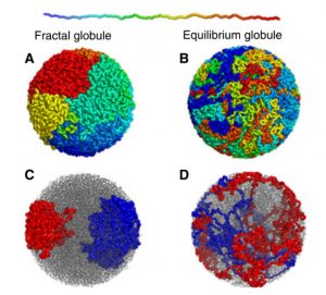
According to current theory, uncoiled chromosomes in the cell nucleus form fractal globules. Unlike the equilibrium globule, which would be the tangled mess of Christmas tree lights, a fractal globule is a maximally compacted structure, but ordered so that a Hamiltonian path is maintained, so called when the thread can be followed through all points without passing through the same point twice, and so that the paths don’t cross, meaning there are no knots. It is an example of the Peano curve, a pattern that can fill an entire plane with a shape that repeats but follows a linear path without crossings. And it adopts a pattern whose structure iterates from small to large scale, as in fractal geometry.
Naturally, this ordered structure is not maintained by itself, but by cellular machinery that untangles knots and breaks crosslinks, such as those that occur when a circular ring of DNA—like bacterial chromosomes—replicates to form two interlocking rings. Topoisomerase enzymes change the topology of DNA to solve these problems: type I enzymes cut and reconnect only one strand of the double-stranded DNA, while type II enzymes cut and reconnect both strands. Other enzymes are also involved in compacting chromosomes for cell division, such as cohesin, a handcuff-like protein that serves to condense the DNA loops and keep the chromatids together, but not scrambled.
Topoisomerases require very fine regulation: if they don’t do their job, the cell dies, but also if there are too many cuts, they irretrievably fragment the DNA. This is why these enzymes are targets for many important drugs, such as antibiotics (quinolones such as ciprofloxacin) that fight bacterial infections and chemotherapy drugs that target cancer. Many of these drugs work not by inhibiting topoisomerases, but by allowing them to act and stabilise DNA breaks so that the cell dies. Some cancer chemotherapies are derived from a natural topoisomerase inhibitor called camptothecin, which is extracted from a tree native to China. Today, molecular modelling allows the design of new chemotherapies that specifically target topoisomerases: mathematics that can save lives.
Comments on this publication